
Optimization of human small intestinal organoids
Collaborating scientists have improved human small intestinal organoids – miniature versions of the small intestine. This will help them to better study the functioning of the small intestine. The scientists from the Hubrecht and AMOLF institutes, and various other collaborators developed organoids that contain mature Paneth cells, which were not present in the earlier human small intestinal organoids. The results of the study were published on 23 August in Cell Stem Cell.
The development of organoids has meant a lot for research on the functioning of human organs and tissues. This has to do with the fact that the mini-organs give a good representation of human biology. However, some of the currently available human organoids, such as the small intestinal organoids, do not yet resemble the original organ fully. Researchers from the Organoid group (Hubrecht Institute) therefore developed a new, optimized version of the miniature small intestines: organoids that include all cell types of the human small intestine.
Missing Paneth cells
The small intestine contains a wide variety of cells, such as enterocytes, stem cells and Paneth cells. Together, these cells form a barrier between the side where the food passes and the side where the blood vessels and immune cells are. The stem cells in the small intestines continuously create all mature cell types, thereby keeping the barrier in good shape. In particular, Paneth cells are important for preventing infections. They do so by producing antimicrobial peptides that act against harmful bacteria. If active Paneth cells are absent, the small intestine will be more prone to infections. This is a problem in several diseases, among which Inflammatory Bowel Disease (IBD). The organoids that used to be employed lacked Paneth cells and did therefore not fully represent healthy human small intestines, while the original mouse organoids described by Toshi Sato in Nature (2009) were complete. This indicated that there was room for improvement.
Surprising finding
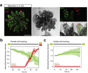
To induce the formation of Paneth cells in human small intestinal organoids, the researchers studied the effect of various molecules. They discovered that the molecule Interleukin-22 (IL-22) increased the number and activity of Paneth cells (Fig. 1a). The effect of IL-22 on Paneth cells is surprising. Researcher Gui-Wei He (Hubrecht Institute) explains why: “At the moment, people believe that IL-22 can promote stem cell function. Our study actually shows that IL-22 does not do this, but rather stimulates the activation of Paneth cells.” The discovered function of IL-22 was therefore used to increase the number of active Paneth cells in human small intestinal organoids. This led to the development of organoids that mimic healthy small intestines.
The origin of the Paneth cells was also a key question of the research. Xuan Zheng (AMOLF): “Cells in the intestine, like in many other organs, are continuously renewed. But how the different cell types emerge in the growing tissue remains incompletely understood, therefore it is advantageous to follow each cell and their type dynamically in time (see figure b).” For this purpose, Zheng developed a method to simultaneously image and track the cell positions within the organoids and also to register the expression of a few key genes that characterize the cell type. Zheng: “While tracking single Paneth cells, I found that they all derived from cells expressing MUC2. MUC2 is a protein involved in forming a mucus layer within the intestine and it characterizes so-called Goblet cells. I saw MUC2 decreasing in some cells, and at the same time there was an increase in the expression of DEFA5, which is an antimicrobial peptide expressed by Paneth cells. Hence, I could directly observe the transformation process of one cell type into another.”
Future directions
Now that optimized human small intestinal organoids are available, researchers can expand on what they study. For instance, they can use the organoids to make mutations: an error in the DNA. Mutations can, among other things, arise if the DNA is copied incorrectly or through external influences. For example, tumor cells often contain mutations in their DNA that are beneficial to their growth. This allows researchers to determine how mutations – occurring in diseases such as IBD – affect the function of the small intestine.
Reference
Gui-Wei He, Lin Lin, Jeff DeMartino, Xuan Zheng, Nadzeya Staliarova, Talya Dayton, Harry Begthel, Willine J. van de Wetering, Eduard Bodewes, Jeroen van Zon, Sander Tans, Carmen Lopez-Iglesias, Peter J. Peters, Wei Wu, Daniel Kotlarz, Christoph Klein, Thanasis Margaritis, Frank Holstege, Hans Clevers, Optimized human intestinal organoid model reveals interleukin-22-dependency of Paneth cell formation, Cell Stem Cell (2022).