Whole-brain imaging in C. elegans
We have developed an imaging experiment capable of simultaneously obtaining ~150 single-neuron Ca2+ activity time series of duration >1hr. The experiment is conducted within a microfluidic device that allows chemosensory stimulus modulation within <1s. These measurements are capable of resolving global activity patterns (correlated dynamics across groups of neurons) on slow (~103 s) timescales, while simultaneously following the activity of key individual neurons such as those involved in sensing (Fig. 1).
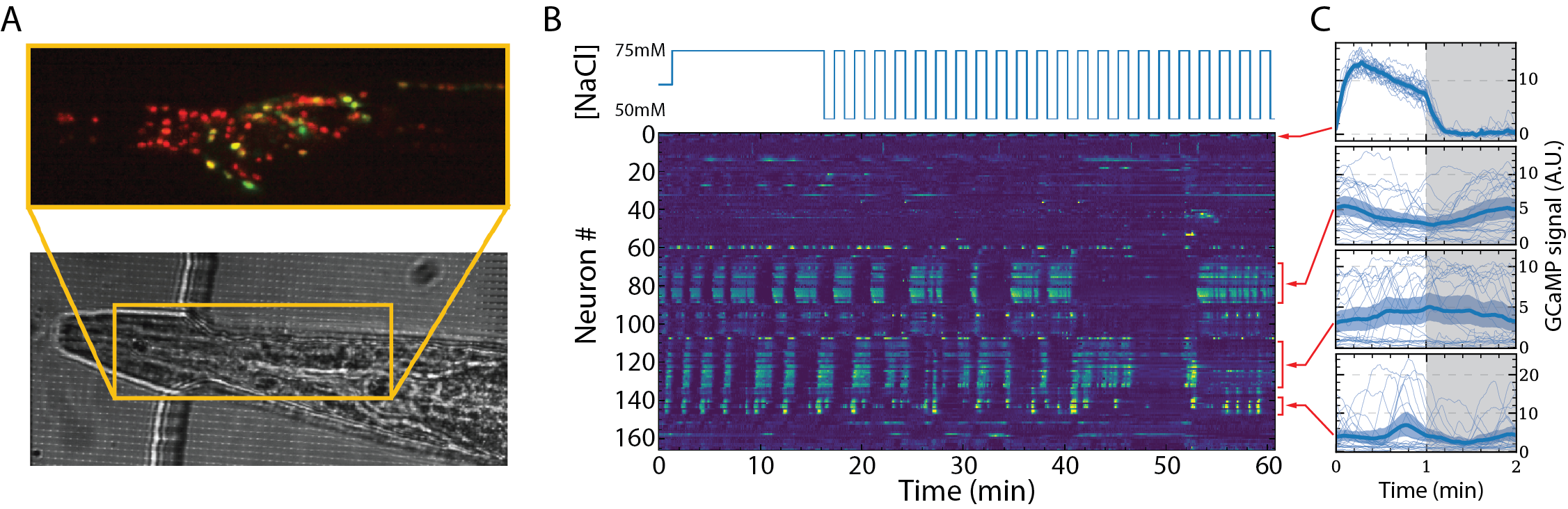
Fig. 1. Resolving whole-brain dynamics at single-cell resolution. (A) The worm is trapped in a narrow channel of a PDMS microfluidic channel (below), with its nose exposed to a flow channel through which chemoeffector stimuli are delivered. (B) Array of Ca2+ activity time series for >160 neurons, shown as a heatmap (bottom) and chemoeffector stimulus time series (top). Large clusters of neurons demonstrate highly correlated oscillatory dynamics. (C) The dynamics of the large clusters (lower three panels) are only weakly correlated with the chemoeffector stimulus level (white=50mM, gray=75mM), whereas a single neuron that a highly deterministic response can be identified.