Spreading focus for better imaging
AMOLF NanoLab news
Extreme Ultraviolet (EUV) light in microscopy offers the advantage of obtaining a high-resolution image combined with spectral information about the object under study. However, because EUV microscopy uses diffraction instead of lenses, imaging with more than one wavelength is challenging. Researchers at ARCNL and Vrije Universiteit Amsterdam have found a work-around by designing and constructing a new class of diffractive optical elements for EUV light. Their results offer possibilities to improve both the light sources and the optical elements in EUV microscopy, paving the way for widespread use of the technique in nanoscience. On January 25th 2021 they published their results in the journal Optica.
EUV microscopy fills the niche between imaging with visible light, which does not provide the nanometer scale detail needed in nanoscience or biological imaging, and imaging methods like electron microscopy, which provides even more detail but is sometimes unsuitable because it needs cryogenic cooling and careful sample preparation. On top of that, due to its strong interaction with matter, EUV light is very useful for spectroscopy measurements revealing material properties of a sample.
However, tabletop EUV microscopy still poses some challenges. “A very practical problem with using EUV light for imaging purposes is that almost every material on earth absorbs most of the radiation. Therefore, we cannot use lenses to focus EUV light”, says ARCNL group leader Stefan Witte. “But, we can use diffraction. If you send light through an object with slits, it will bend. If the slits are arranged the right way, it is possible to focus the radiation, just like you would focus visible light with a lens.”
Zoneplates instead of lenses
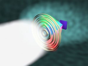
EUV light can be focused with a so-called Fresnel zoneplate, a disc with a circular pattern of slits that diffracts the light. An inherent property of diffraction, however, is that the angle of diffraction is dependent on the wavelength. Witte: “We use a coherent source that contains a broad spectrum of light in the EUV range. With a conventional zoneplate, this results in different focus points for each wavelength in the beam, but we can only use one of them without having to move the sample. Moreover, it is impossible to collect spectral data of a sample when you only send one wavelength of light through it. The material properties of the sample that we could unveil with EUV spectroscopy thus stay hidden.”
Optimization
Lars Loetgering and Kevin Liu, both scientists in Witte’s group, found a somewhat counterintuitive solution to this problem. While a perfect zoneplate offers distinct focus points, minor faults or irregularities in the circular pattern of slits will cause the focus to get smeared in the direction of the beam. The researchers realized that they could use these messy focus ‘smears’ to their advantage. “The focus smears are also shifted for each wavelength in the spectrum, but they do overlap somewhat”, says Witte. We made a model to calculate the optimum zoneplate, in which a minimum of irregularities – or entropy in the structure – results in a maximum overlap of the focus smears. With that, we can get the most out of the available EUV light and also take advantage of the spectral sensitivity of EUV imaging by collecting the data from up to nine different wavelengths.”
NanoLab AMOLF
Using the NanoLab facilities at AMOLF, Loetgering and Liu got to work to construct their imperfect zoneplate. First, they used the Leica sputter coater to deposit a 5 nanometer chromium adhesion layer on a 50 nanometer thick free standing Si3N4 membrane. After this, a 90 nanometer thick gold layer was sputter coated on top. The final step involved milling the zoneplate on this substrate, for which Loetering and Liu used the FEI Helios Nanolab 600 that combines a scanning microscope (SEM) and gallium focused ion beam (FIB). This combination is convenient because it allowed them to write patterns on the substrate and simultaneously monitor their progress. Looking back at the work done at AMOLF NanoLab, Liu says: “it is a great facility with a large collection of state-of-the-art equipment that is available to researchers from all over the Netherlands. It allows them to realize their most novel and innovative research ideas without having to outsource tasks. I think this is quite unique.”
Exciting times ahead
Witte and his team have tested their ‘imperfect’ zoneplates both in simulations and experiments and are excited about the results. “This new type of diffractive optical elements is not only paving the way towards widespread use of tabletop EUV microscopy, but we can also use it to take a step back and try to make our EUV sources more efficient”, he says. “We are looking for the ideal combination of light and diffraction, which can be different depending on the information you are searching for.”
Witte expects the years ahead to be crucial for a wider use of EUV microscopy in nanoscience; “the technique is currently limited by the efficiency of the sources and the restriction to single wavelength radiation. There is still a lot of work to be done, but with our approach I expect we can optimize the technique further so that it can be used in metrology or materials science. For example, researchers who are now dependent on large synchrotron facilities will be able to do their experiments in their own lab with a tabletop EUV microscope.”
Reference
Lars Loetgering, Xiaomeng Liu, Anne C. C. De Beurs, Mengqi Du, Guido Kuijper, Kjeld S. E. Eikema, and Stefan Witte, Extreme ultraviolet multispectral ptychography with minimum entropy beams, Optica, 8 (2), 130-138 (2021).