Symposium on Light-Driven Processes Monitored Inside the TEM : Keynote Speakers
Sophie Meuret
Exploring the Dynamics of Semiconductors with an Ultrafast Transmission Electron Microscope
In this presentation, for example, we will discuss the advantage and inconvenient of TRCL in a UTEM and present our results on the study of charge carrier dynamics in a 4 nm In0.3Ga0.7N/GaN quantum well. We studied the QW emission dynamic both along and across the quantum well and correlate the results with strain map and high resolution HADF images.
References
[1] P. Corfdir et al., “Exciton localization on basal stacking faults in a-plane epitaxial lateral overgrown GaN grown by hydride vapor phase epitaxy,” J. Appl. Phys., vol. 105, no. 4, p. 043102, 2009.
[2] R. J. Moerland, I. G. C. Weppelman, M. W. H. Garming, P. Kruit, and J. P. Hoogenboom, “Time-resolved cathodoluminescence microscopy with sub-nanosecond beam blanking for direct evaluation of the local density of states,” Opt. Express, vol. 24, no. 21, p. 24760, 2016.
[3] X. Fu et al., “Exciton Drift in Semiconductors under Uniform Strain Gradients: Application to Bent ZnO Microwires,” ACS Nano, vol. 8, no. 4, pp. 3412–3420, 2014.
[4] S. Meuret et al., “Time-resolved cathodoluminescence in an ultrafast transmission electron microscope,” Appl. Phys. Lett., vol. 119, no. 6, p. 6, 2021.
[5] Y. J. Kim and O. H. Kwon, “Cathodoluminescence in Ultrafast Electron Microscopy,” ACS Nano, vol. 15, no. 12, pp. 19480–19489, 2021.
[6] F. Houdellier, G. M. Caruso, S. Weber, M. Kociak, and A. Arbouet, “Development of a high brightness ultrafast Transmission Electron Microscope based on a laser-driven cold field emission source,” Ultramicroscopy, vol. 186, pp. 128–138, 2018.
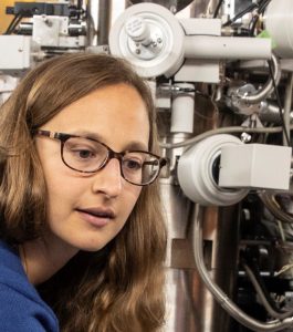
Sophie Meuret: My research activities focus on the optical properties of nano-objects, which I study by combining experimental, instrumental and theoretical approaches. More specifically, I am interested in the study of nano-optical materials e.g. quantum wells, point defects in diamond or boron nitride, plasmonic and semiconductor nanostructures. The guiding thread of my research is based on understanding and exploiting the interaction of electrons (or light) with matter using light (or electrons). To do so, I have studied the optical properties of materials with cathodoluminescence (CL), using different types of scanning electron microscopes, with various characteristics ranging from nanometer spatial resolution to picosecond temporal resolution. As an independent researcher at CEMES, I am developing new time-resolved electron-based spectroscopy techniques for nano-optics developing mostly photon near-field electron microscopy (PINEM) and time-resolved CL to study the dynamics of semiconductors.
Armin Feist
Merging Electron Microscopy with Advanced Photonics
My talk will discuss recent progress in coupling free electrons and photons via inelastic scattering for the optical mode- and phase-resolved analysis of plasmonic nanocavities. Furthermore, stimulated interaction at an integrated photonic microresonator enables continuous-wave optical phase modulation, while spontaneous scattering produces electron-photon pair states. In analogy to spontaneous parametric down-conversion, this mechanism promises heralded single-electron or Fock-state photon sources.
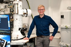
Dr. Armin Feist is a scientist in the Department of Ultrafast Dynamics at the Max-Planck Institute for Multidisciplinary Sciences. After studying in Leipzig, Leeds, and Göttingen, his Ph.D., working in the group of Prof. Claus Ropers at the University of Göttingen, focused on developing and applying Ultrafast TEM using coherent electron pulses. His distinctions include the 2019 EPS-QEOD Thesis Prizes for applied aspects and the Optica Li Innovation Prize 2022. Current research interests are nanoscale structural dynamics, ultrafast plasmonics, and optically tailored free-electron beams. This entails exploring new instrumental capabilities in electron microscopy, with the vision of combining ultrafast optics and integrated photonics with state-of-the-art electron microscopes.
Ulrich Lorenz
Observing Nanoscale Dynamics with Time-Resolved Electron Microscopy
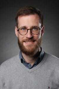
Ulrich Lorenz: After a Ph.D. in molecular physics, Ulrich Lorenz did a postdoc with Ahmed Zewail at Caltech, where he became interested in time-resolved and ultrafast electron microscopy. Since 2016, he has been an Assistant Professor at EPFL in Lausanne, Switzerland, where his research has been supported by an ERC Starting Grant, and most recently by a Consolidator Grant of the Swiss National Science Foundation. His interests revolve around the development of new methods to image the dynamics of nanoscale systems at high spatial and temporal resolution. Recently, his group has introduced a new approach to time-resolved cryo-electron microscopy that enables the observation of protein dynamics with microsecond time resolution.
Jennifer Dionne
The Light Stuff: Enabling Sustainable Chemical Manufacturing with Atomically-Optimized Photocatalysts
Here, we present our research advancing plasmon photocatalysis from the atomic to the reactor scale. First, we describe advances in in-situ atomic-scale catalyst characterization, using environmental optically-coupled transmission electron microscopy. With both light and reactive gases introduced into the column of an electron microscope, we can monitor chemical transformations under various illumination conditions, gaseous environments, and at controlled temperatures, correlating three-dimensional atomic-scale catalyst structure with photo-chemical reactivity. Then, we describe how these atomic-scale insights enable optimized reactor-scale performance. As model systems, we consider three reactions: 1) acetylene hydrogenation with Ag-Pd catalysts; 2) CO2 reduction with Au-Pd catalysts; and 3) nitrogen fixation with AuRu catalysts. Here, Au/Ag acts as a strong plasmonic light absorber while Pd/Ru serves as the catalyst. We find that plasmons modify the rate of distinct reaction steps differently and that reaction nucleation occurs at electromagnetic hot-spots – even when those hot-spots do not occur in the preferred nucleation site. Plasmons also open new reaction pathways that are not observed without illumination, enabling both high-efficiency and selective catalysis with tuned bimetallic catalyst composition. Our results provide a roadmap for how atomically-architected photocatalysts can precisely control molecular interactions for high-efficiency and product-selective chemistry.
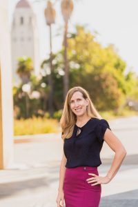
Jennifer (Jen) Dionne is a Professor of Materials Science and Engineering and, by courtesy, of Radiology at Stanford. She is also a Chan Zuckerberg Biohub Investigator, deputy director of Q-NEXT (a DOE-funded National Quantum Initiative), and co-founder of Pumpkinseed, a company that directly sequences peptides for improved immunotherapies. From 2020-2023, Jen served as Stanford’s Inaugural Vice Provost of Shared Facilities, raising capital to modernize instrumentation, fund experiential education, and support new and existing users of the shared facilities. Jen received her B.S. degrees in Physics and Systems Science and Mathematics from Washington University in St. Louis, her Ph. D. in Applied Physics at the California Institute of Technology in 2009, and her postdoctoral training in Chemistry at Berkeley. As a pioneer of nanophotonics, she is passionate about developing methods to observe and control chemical and biological processes as they unfold with nanometer scale resolution, emphasizing critical challenges in global health and sustainability. Her research has developed culture-free methods to detect pathogens and their antibiotic susceptibility; amplification-free methods to detect and sequence nucleic acids and proteins; and new methods to image light-driven chemical reactions with atomic-scale resolution. Her work has been recognized with the Alan T. Waterman Award, a NIH Director’s New Innovator Award, a Moore Inventor Fellowship, the Materials Research Society Young Investigator Award, and the Presidential Early Career Award for Scientists and Engineers, and was featured on Oprah’s list of “50 Things that will make you say ‘Wow’!”. She is especially proud of the many outstanding alumni who have graduated from her lab. Dionne lab members have received awards including HHMI, Beckman, Schmidt and Packard fellowships, and alum now hold faculty positions spanning top academia (eg, professors at MIT, Stanford, Berkeley, Northwestern, among others), industry (Apple, Intel, Facebook/Meta, Lockheed Martin, Pacific Biosciences, Tempus), startups (founders of Antora and Arabesque), policy (Congressional Fellows), and communications (including a Pulitzer prize winner). She also perceives outreach as a critical component of her role and frequently collaborates with visual and performing artists to convey the beauty of science to the broader public. Beyond the lab, Jen enjoys long-distance cycling, trail running, and reliving her childhood with her two young sons.